Eyes - Don’t lose sight of Glaucoma
Updated: 4/8/24
Information on primary angle closure glaucoma in Bouviers des Flandres
Information on primary angle closure glaucoma in Bouviers des Flandres
|
What is glaucoma?
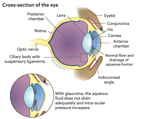
Glaucoma is an eye disease where aqueous humor builds up, increasing the eye's intraocular pressure (IOP). This increased pressure will damage the optic nerve and can lead to blindness. What are the essential eye structures in glaucoma? Anterior chamber, Cornea, Iris, Iridocorneal angle, Ciliary cleft, and Optic nerve How is glaucoma classified? It is called primary when the fluid buildup is linked to genetic abnormalities in the drainage pathway or secondary when another ocular disease (e.g., lens luxation, uveitis) is present. Also, glaucoma can be classified according to the state of the drainage angle. The angle may be open (in which case the obstruction is further downstream), narrow, or closed. What causes the Intraocular pressure to increase? Aqueous humor is continuously being produced. It contains nutrients and oxygen used within the eye. As new fluid is made, the old fluid exits the eye through the drainage angle (between the cornea and iris). The drainage angle is also called the iridocorneal angle. The ciliary cleft and pectinate ligaments are vital structures in the drainage angle. Blockage of the drainage pathway allows for increased IOP. Iridocorneal angles are classified as open or narrow/closed. What is primary angle closure glaucoma (PACG) in Bouviers? PACG is genetic. The dog inherits abnormalities of the drainage pathway; in other words, the pathway angles are narrow or closed, interfering with the draining of the aqueous fluid. In addition, bouviers are known to have pectinate ligament dysplasia (PLD), an inherited abnormality that is a factor in the narrowing of the drainage angles. However, only a small proportion of dogs with PLD will develop PACG in their lifetime. How do you test for primary angle closure glaucoma? Two tests are commonly done. The first is tonometry. This test is for IOP. The second is a gonioscopy test. This test is used to evaluate the drainage angles. Dogs with narrow angles and other risk factors, such as a history of glaucoma in a close relative ( litter mate, parents, or grandparent ), are at risk of developing PACG. Therefore, they should have an additional test called ultrasound biomicroscopy (UBM). This test provides more detailed information on the drainage pathway, particularly evaluating the ciliary cleft. Eye Angles are not static. They change as the dog ages and must be monitored over the dog's life. What are the risk factors for developing PACG
What are the symptoms of primary angle closure glaucoma (PACG)
How can primary angle closure glaucoma (PACG) be treated? Obtaining medical care as quickly as possible is essential to reduce the risk of irreversible damage and blindness.
What can be done to protect your dog from vision loss?
What is the role of the responsible dog breeder?
|

